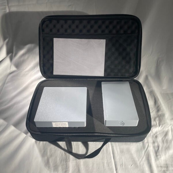
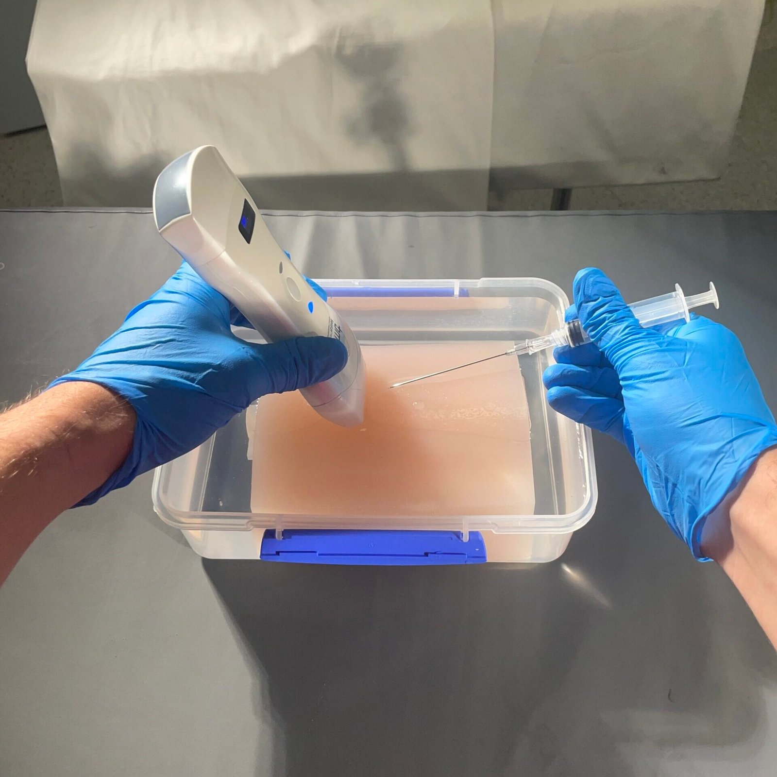
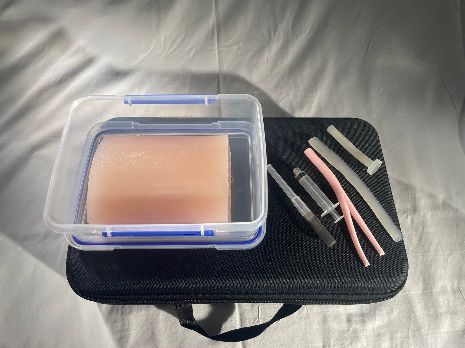
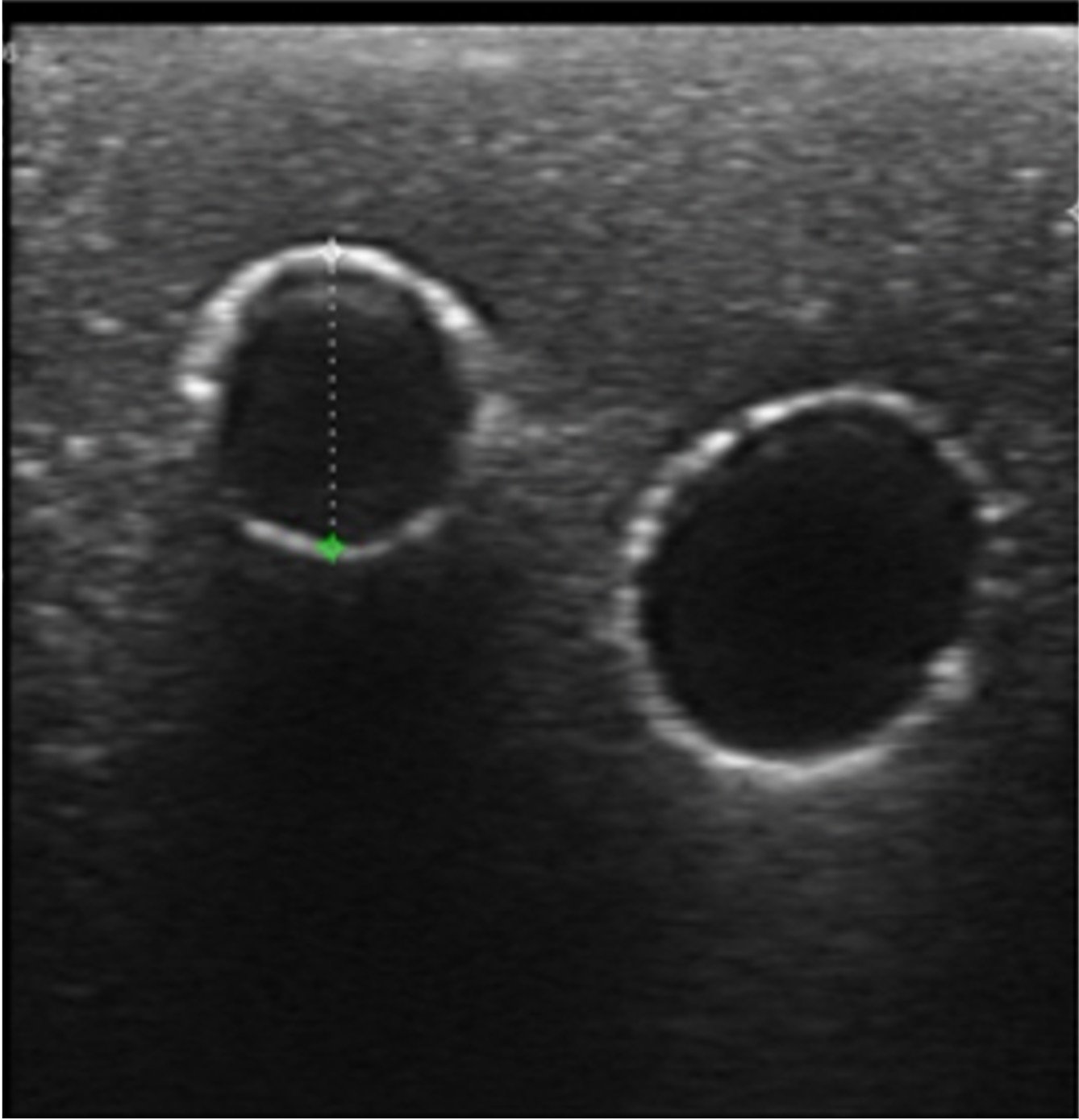
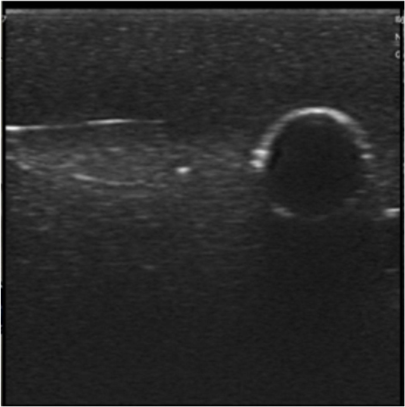
Clinically validated for ultrasound-guided femoral vascular access training, this reusable phantom reproduces the femoral nerve, common femoral vein and common femoral artery with bifurcating branches. Open-ended vessels support needle insertion, guidewire passage and external flow systems, while the water-filled, self-healing elastomer design enables repeatable training and experimental work with minimal consumables.
| WHAT CAN BE PRACTICED
• Ultrasound-guided femoral arterial and venous access • Nerve block • Vessel compression and haemostasis technique |
TRAINING SETTINGS
• Classroom/office • Clinical centres • Research laboratories |
EDUCATIONAL VALUE
• Realistic repeatable anatomy without patient exposure • Realistic tactile and ultrasound response • Self-healing design reduces consumable use |
| TECHNICAL SPECIFICATIONS
Needle sizes: 18-23G bevelled needles Puncture density: up to 1 isolated puncture/mm² Self-healing: 18-20G tracks heal within 3 h; unapparent by 24 h Sound speed: 1454 ± 15 m/s Shore hardness: OO 18.0 ± 2.2 Colours: Light, Medium, Dark, Clear |
INTENDED TRAINING APPLICATION
• Femoral arterial access training • Femoral venous access training • Ultrasound needle visualisation and targeting • Needle-to-guidewire access sequence • Vessel compression and haemostasis demonstration • Flow-system, protocol and device testing |
USAGE AND LIFESPAN
| ~2,000
MAX. ESTIMATED PUNCTURES Across 4x5cm region of inetrest at 1 puncture/mm². |
3 h
SELF-HEALING 18G tracks heal under ultrasound within 3 h. |
24 h TOTAL RECOVERY 18G tracks became fully unapparent within a day. |
CLINICALLY VALIDATED
Peer-reviewed evaluation published in Applied Sciences assessed the simulator with 18 undergraduate medical students during ultrasound-guided femoral vascular access training and 31 medical educators using a seven-question Likert survey. After only 5 minutes of free practice, mean vascular access time improved from 107 s to 44 s, with most students experiencing a time improvement of more than 60%. Educator feedback was also strongly positive, with most respondents reporting that they would adopt the phantom for teaching.
| 60% IMPROVEMENT
IN OVER 50% OF STUDENTS Timed before and after 5 min of free practice. |
107→44 s
MEAN ACCESS TIME Average procedural time reduced by ~59%. |
31/31 EDUCATORS Reported they would use the phantom for training. |
Manuals and Technical Sheets:
Technical Sheet – Click here to download the Technical Sheet